Application Focus - Electroporation for High-Efficiency Transformation of Chlamydomonas
By Michelle M. Ng-Almada, Ph. D.

In this blog we will first overview the model system Chlamydomonas reinhardtii and methods for transforming this single celled, flagellated alga. We will provide protocol recommendations for high-efficiency square wave electroporation with generators such as the ECM 830 which can achieve a transformation efficiency of 2 to 6 x 10³ transformants per µg of exogenous DNA. Next, we will cover protocol recommendations for exponential decay wave generators, such as the ECM 630, which can achieve transformation efficiencies of 2 to 3 x 10³ transformants per µg of exogenous DNA. Finally, we will compare the pros and cons and typical efficiencies of these methods to traditional glass bead transformation and electroporation via decaying square wave generators.
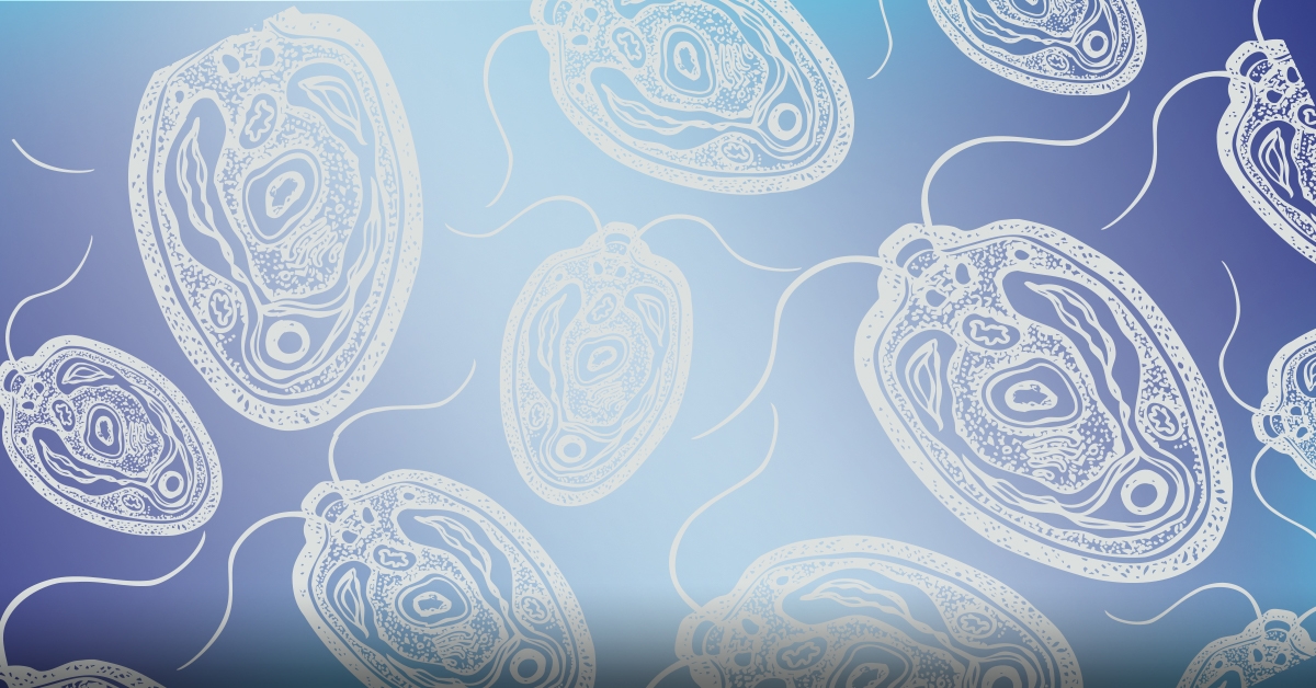
Chlamydomonas reinhardtii is an excellent model system for studying basic biological structures and processes
- Chlamydomonas has two flagella which are similar in structure and function to cilia in other organisms. These flagella can be isolated for biochemical and molecular analysis, and have been used for many years as a model system to study cilia biogenesis and intraflagellar transport (IFT).
- Chlamydomonas may be used to study photosynthesis. It can grow on simple salt medium, utilizing photosynthesis to derive energy, or can grow in comlet darkness if acetate is provided as an alternate carbon source.
- C. reinhardtii exists in a haploid vegetative state, or under adverse environmental conditions the two mating types may fuse and create a diploid zygospore. When the conditions improve, the diploid zygote undergoes meiosis and releases four haploid cells that resume the vegetative life cycle.
- The full sequences of the nuclear, chloroplast, and mitochondrial genomes of C. reinhartdii have been sequenced and mapped.
- These characteristics allow for both forward and reverse genetic screening techniques.
- Additionally, the Chlamydomonas Resource Center offers a repository for wild type and mutant cultures and reagents.
- Electroporation is an efficient, convenient method of introducing exogenous DNA without having to remove the Chlamydomonas cell wall.
High-Efficiency Square Wave Electroporation Protocol
- Grow cells in liquid TAP medium to a cell density of 1 to 2 x 10⁷ cells/ml.
- Inoculate cell stock into fresh liquid TAP medium to a concentration of 1 x 10⁶ cells/ml and grow under continuous light for 18 to 20 hours until a cell density of 4 x 10⁶ cells/ml is reached.
- Centrifuge cells at 1250 g for 5 minutes at room temperature, wash, and resuspend with pre-chilled TAP medium containing 60 mM sorbitol, and put the mixture on ice for 10 minutes.
- Place 250 µl of cell suspension (or 5 x 10⁷ cells) into a pre-chilled 4 mm gap electroporation cuvette, and add 100 ng of desired exogenous DNA.
- Electroporate with a generator with square wave form, such as ECM 830 or Gemini X2, using the following electroporation parameters: 500 V, 4 ms pulse duration, 6 to 7 pulses, 100 ms pulse interval time.
- Immediately place the cuvette on ice for 10 minutes after electroporation, then transfer cell suspension into a 50 ml conical centrifuge tube containing 10 ml TAP medium to recover overnight with dim light and slow shaking.
- Plate cells as desired with appropriate selection medium.
- Expected transformation efficiency is 2 to 6 x 10³ transformants per µg of exogenous DNA.
- Please see Wang L. et al. 20191 for protocol details.
Exponential Decay Wave Electroporation Protocol
- Prepare cells pre- and post- electroporation as described above for Square Wave Electroporation Protocol
- Electroporate with a generator that has exponential decay waveform, such as ECM 630 or Gemini X2, using the following electroporation parameters: 800 V, 1575 Ω resistance, 50 µF capacitance.
- Desired pulse length is 10 to 14 ms; above or below this desired pulse length range may reduce transformation efficiency.
- When tested side by side with above high-efficiency square wave electroporation, this exponential decay wave method yielded approximately 50% of the efficiency of square wave.
- Please see Wang L., et al. 20132 for protocol details.
Comparison with other Chlamydomonas transformation methods
- Glass bead transformation3,4 utilizes agitation with glass beads to introduce exogenous DNA. This method does not require special equipment, however it requires the use of cell-wall deficient strains for removal of cell wall prior to the glass bead agitation. When compared side by side with the above high-efficiency square wave electroporation method, glass bead method was only about 20% of the efficiency of the square wave method.
- Electroporation with another type of electroporator (decaying square wave pulse generator NEPA21), like the square wave and exponential decay wave electroporation methods described above, has the benefit not requiring cell wall removal. Transformation with this type of electroporator is reported to yield 0.4 to 3 x 10³ transformants per µg of DNA5. However this method requires a relatively large amount of exogenous DNA (400 µg per reaction) which is not ideal for genetic screening applications.
- In comparison, high-efficiency square wave electroporation and optimized exponential decay wave electroporation with generators such as ECM 830 and ECM 630 yield transformation efficiencies of up to 6 x 10³ transformants per µg of exogenous DNA, does not require removal of cell walls, and requires only 100 µg of exogenous DNA per transformation reaction.
References:
1. Wang, L., et al. (2019) Rapid and high efficiency transformation of Chlamydomonas reinhardtii by square-wave electroporation. Biosci Rep 39 (1): BSR20181210.
2. Wang, L., et al. (2013) Flagellar regeneration requires cytoplasmic microtubule depolymerization and kinesin-13. J. Cell Sci. 126, 1531–1540.
3. Kindle, K.L. (1990) High-frequency nuclear transformation of Chlamydomonas reinhardtii. Proc. Natl. Acad. Sci. USA 87, 1228–1232.
4. Nelson J.A. and Lefebvre P.A. (1995) Transformation of Chlamydomonas reinhardtii. Methods Cell Biol. 47, 513–51.
5. Yamano T., Iguchi H. and Fukuzawa H. (2013) Rapid transformation of Chlamydomonas reinhardtii without cell-wall removal. J. Biosci. Bioeng. 115, 691–694.
Buy an ECM 830 Electroporation System, get a free BTXpress High Performance Electroporation Buffer—Click here for promotion details!


 800-272-2775
