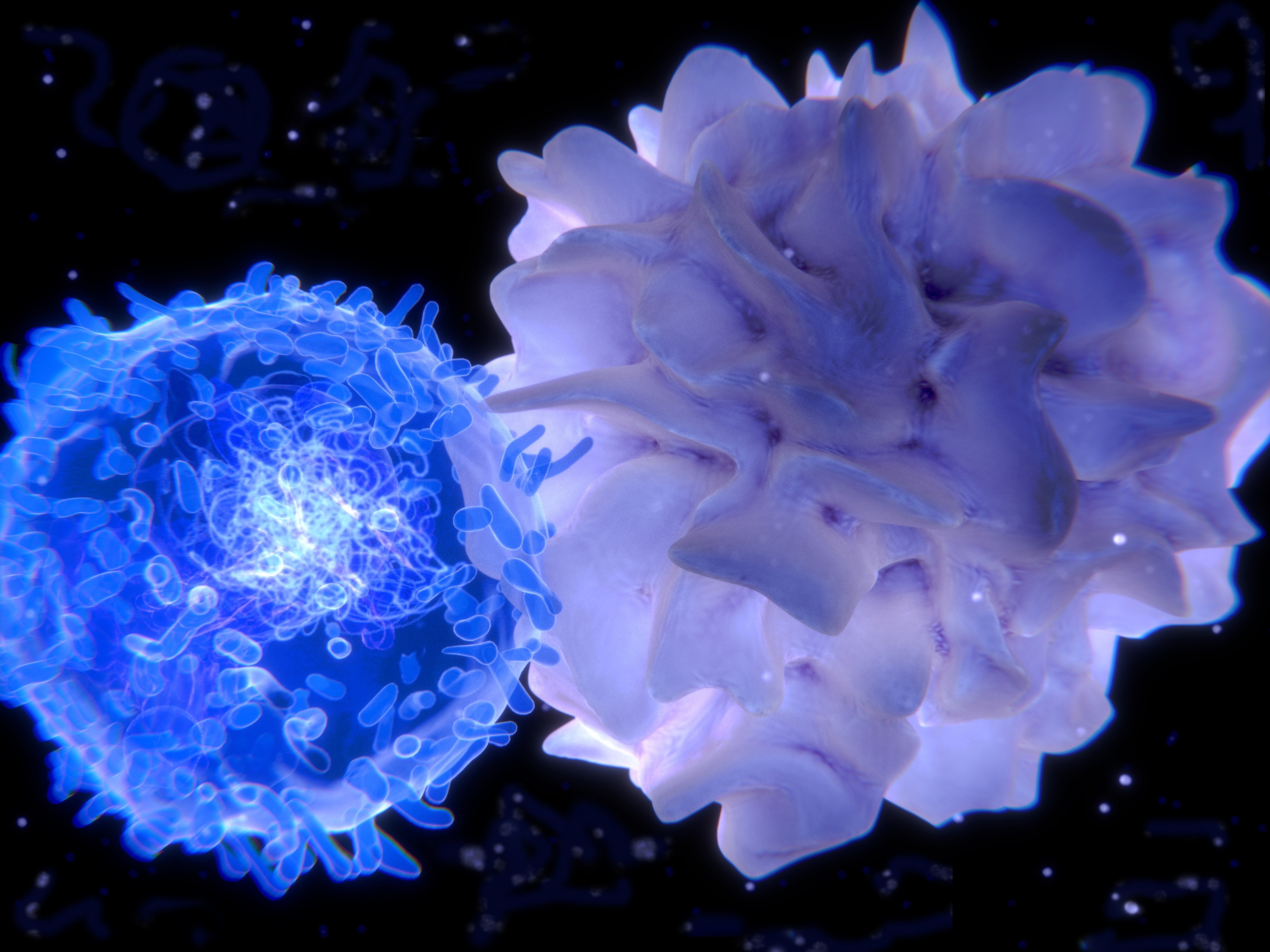
Application Focus - Modeling Dendritic Cell and T-Cell Interactions in a 3D Environment
By George Kamphaus, Ph. D.
 This blog describes a novel system for the study of T-cell activation by antigen-presenting dendritic cells (DC) and the use of a BTX ECM 830 electroporator to transfect quiescent (naïve or memory) T-cells. While there are many T-cell therapies that use genetically modified or transfected T-cells, in nearly all cases, the T-cells are activated before transfection. Such methods preclude the study of the physiologic mechanisms of activation by DC cells. To increase the understanding of the intercellular dynamics of quiescent CD8+ T-cell interaction with DC and CD-4+ helper cells, Abu Shah, et. al. have created a 3-D system that reconstitutes a tissue-like environment that allows for the integration of cytokines, DC and quiescent T-cells. Utilizing electroporation to transfect mRNA constructs of modified antigen-specific TCRs, the researchers were able to express functional TCR on the surface of the quiescent T-cells and show activation by interaction with DCs loaded with the corresponding peptide antigen.
- Harvest and wash T-cells three times with Opti-MEM (Thermo Fisher Scientific). Resuspend cells at 25 x 106 cells/ml.
- Aliquot 100 to 200 µl (2.5 to 5 x 10⁶ cells) into separate tubes and mix with the desired mRNA products.
- For 106 CD8 T-cells, use 2 µg of each TCRα, TCRβ and CD3ζ RNA. For 106 CD4 T-cells, use 4 µg of TCRα, TCRβ RNA.
- Add mixture to electroporation cuvette (Cuvette Plus, 2 mm gap, BTX).
- Electroporation Parameters for the BTX ECM 830: 1 pulse (square wave) at 300 V, 2 ms pulse duration.
- Collect cells from the cuvette and culture in 1 ml of pre-warmed media.
- Labeling with different fluorescent markers allows different cell types to be monitored simultaneously by time-lapse video. In this study the motility of CD4+ and CD8+ T-cells and DC were all monitored together in a single system under identical conditions.
- Different chemokines can be added to the matrix.
- T-cells can be added before or after solidification of the collagen matrix to study varying kinetics of the DC / T-cell presentation.
- The effect of TCR-antigen affinity can be studied by expression of various TCR in quiescent T-cells.
- This can be used as a model system for immune-modulation therapies, such as PD-1 checkpoint inhibitors.
- Because cells can be easily extracted after observation, cell surface markers and other characteristics can be studied by FACS or other flow cytometry methods.
- Using the BTX ECM 830 square wave electroporator, the efficiency of TCR expression varied among donors with an average value of 81 ± 7% (mean ± SD, n = 13), with more than 90% cell viability and 80–90% cell recovery, both of which were severely reduced using alternative electroporation approaches (Amaxa and Neon).
- The induction of expression of an exogenous TCR in CD4 T-cells is considerably harder than in CD8 T-cells (Dai et al., 2009). Using natural sequences or the introduction of cysteine modifications used for 1G4 and 868 failed to induce expression of TCRs in naïve CD4 T-cells.
- Cell motility of both CD4+ and CD8+ T-cells was faster than DC, either with or without homeostatic chemokines.
- While both high affinity and low affinity peptide-TCR interactions were able to activate CD8+ T-cells, as evidenced by CD69 and CD25 expression, only T-cells with high-affinity TCR-antigen interactions showed arrested motility.
- Two different anti-PD-1 antibodies enhanced the clustering of CD-8 T-cells around their targets, while not changing the motility dynamics.
- Using the BTX ECM 830 square wave electroporator, the cells maintained the motile behavior and interaction dynamics of naïve CD8 T-cells expressing 1G4, at similar levels to those of untouched cells, on 2D stimulatory ‘spots.'
Reference:
1. Abu-Shah, E., et al. A tissue-like platform for studying engineered quiescent human T-cells' interactions with dendritic cells. eLife 2019; 8.
Click here to visit our Protocol Database, for electroporation protocols searchable by, system, cell/tissue type, application/transfectant, and citation.


 800-272-2775
